
Bioguard Corporation
Encephalitozoon cuniculi is a microsporidial, unicellular, spore-forming, obligate intracellular parasite. It can invade the host’s central nervous system, kidneys, crystals, etc. E. cuniculi affects rabbits by causing damage to the brain, nervous system, kidneys, and other important organs. E. cuniculi pose a zoonotic risk to immune-compromised humans. In addition, it can infect various mammals, such as rabbits, rats, mice, horses, foxes, cats, dogs, muskrats, leopards, and baboons.
Transmission and Life Cycle
E. cuniculi has a direct life cycle with both horizontal and vertical (transplacental) transmission. In rabbits, the common routes of natural horizontal infection are via the ingestion of contaminated food or water or, less commonly, via inhalation of spores.
After ingestion, the spores invade enterocytes and then spread through bloodstream or the lymphatic system. Then, it is carried into the blood circulation to target organs (kidney, central nervous system, eye, liver, and heart) where it causes inflammation. Antibody can be detected 2-3 weeks after infection, and IgM are usually detectable up to 18 weeks post-exposure. Spores are passed in the urine of rabbits, beginning around 35 days after infection, and continue to be excreted for 2 to 3 months.
Serological surveys show high seroprevalence rates (23-75%) of E. cuniculi in rabbits. The seroprevalence rate of E. cuniculi is about 63.2-67.8% in Taiwan. However, most are asymptomatic and few are found to be affected by disease.
Clinical signs
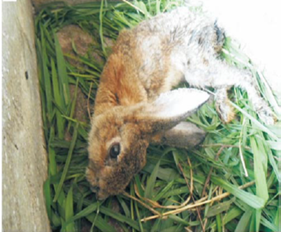
When E. cuniculi infect the rabbits, the common clinical signs include head tilt (vestibular disease), hind limb paresis (weakness of the hind limbs), urinary incontinence, renal failure, cloudy eyes (anterior uveitis), cloudy lenses of the eyes (cataracts), or even blindness.
Diagnosis
Clinical diagnosis of encephalitozoonosis can be challenging because of the following reasons. 1. Serologic evidence is strong evidence of infection but not indicative of clinical signs. 2. Seroconversion does not result in a protective response for the patient. 3. Histologic severity and distribution of lesions are not directly correlated with the severity of clinical signs. 4. Most infected rabbits are asymptomatic or carriers. Diagnostic methods of encephalitozoonosis include histopathology, serology, and molecular diagnosis.
• Histopathology- histological examination combined with special staining, or concentrated urine for cytological microsporidia detection
• Serology- indirect immune fluorescence antibody test, direct agglutination test, ELISA, western blot
• Molecular diagnosis- PCR
Treatment and Prevention
No uniformly effective treatment has been established. Fenbendazole, Albendazole, and Oxibendazole may be effective in vivo. The treatment of choice generally is fenbendazole, because it has been shown to both prevent and treat E cuniculi infections. To reduce the inflammatory reaction, steroids or NSAIDS may be applied. The use of systemic steroid therapy, although controversial, has been advocated to reduce severe CNS inflammation. The risk with this treatment is that corticosteroids might suppress the immune system to the point that E cuniculi or other infectious organisms may create additional problems. Cases involving the ocular form need to be referred to a veterinary ophthalmologist if surgery is recommended. Prevention of the disease from spreading includes thoroughly cleaning the rabbit’s environment, applying a diluted solution of bleach (1:32) for disinfection, and providing clean water and food to rabbits.
