

Bioguard Corporation
The coronavirus of ferrets was first described in 1993. This ferret enteric coronavirus (FRECV) caused an enteric disease called epizootic catarrhal enteritis (ECE) or green slime disease. Later, a ferret systemic coronavirus (FRSCV)-associated disease, resembling the dry form of feline infectious peritonitis (FIP), was identified. ECE is characterized by high morbidity (nearly 100%) but low mortality (< 5%). FRSCV is associated with a high mortality rate and short duration of illness.
Pathogen
Coronaviruses are large, enveloped, positive-stranded RNA viruses classified under the genus Coronavirus within the family Coronaviridae, order Nidovirales. Virions are roughly spherical and are notable for the large spike (S) glycoprotein that extends from the virus envelope, which resembles a crown or solar corona when imaged using an electron microscope. The diameter of ferret coronavirus is about 120 nanometers, and its genome is about 28 kilobases.
Transmission
Both the enteric (FRECV) and systemic (FRSCV) ferret coronaviruses are classified as group 1 coronaviruses (that is, alphacoronaviruses), related to feline coronavirus and canine enteric coronavirus. The routes for transmission of FRECV and the FRSCV are suggested to be fecal–oral. Many facts of the pathogenesis of the virulent systemic ferret coronavirus remain unknown, but similar to FIPV, macrophages appear to play an important role in the inflammatory response.
Clinical signs
After infection of FRECV, the virus causes blunting of the intestinal villi and consequent maldigestion and malabsorption. Clinical signs include anorexia, vomiting, diarrhea, melena, dehydration, lethargy, and weight loss. The microscopic lesions include diffuse lymphocytic enteritis, with villus atrophy, fusion, and blunting; vacuolar degeneration and necrosis of the apical epithelium; or a combination of all these lesions.
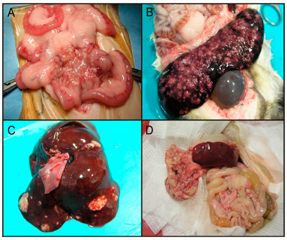
FRSCV-associated disease causing FIP-like lesions has been reported in mostly young (<18 months) ferrets. Clinical signs are nonspecific, including anorexia, weight loss, diarrhea, and enlarged intra-abdominal and, less commonly, peripheral lymph nodes. FRSCV is grossly associated with pale to white nodules (granulomatous inflammation) in multiple organs, including the spleen, kidneys, mesenteric lymph nodes, intestines, liver, lungs, and brains. Granulomas have a heterogenous cellular composition including macrophages, T and B lymphocytes, and plasma cells.
Diagnosis
Definitive diagnosis requires intestinal biopsy and histopathological examination, with confirmation of viral antigen or nucleic acid by immunohistochemistry or in situ hybridization. RT-PCR and electron microscopy can be used to examine feces from ferrets. Systemic coronavirus disease in ferrets can be confirmed by histological evaluation of biopsies and intralesional coronavirus nucleic acid detection.
RT-PCR has been used to identify viruses in tissues and can differentiate ferret enteric coronaviruses from ferret systemic coronaviruses.
References
