
Bioguard Corporation
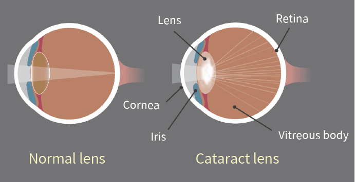
Canine juvenile cataract (JHC) is a hereditary form of cataract characterized by cloudiness and degeneration of the lens in the eye. This condition prevents light and images from passing through the lens to the retina, impairing the dog’s vision and eventually leading to blindness. Affected dogs may exhibit symptoms such as bumping into furniture, losing direction, and moving slowly. JHC can also lead to intraocular complications, including uveitis, glaucoma, and lens luxation. Dogs are more prone to cataracts than any other species, and while cataracts can develop at any age, juvenile cataracts typically occur in dogs under 7 years old.
Genetic mutaion related to JHC
JHC is caused by a mutation in the heat shock transcription factor 4 (HSF4) gene, leading to an autosomal recessive inheritance pattern. Assuming that “A” represents a normal allele and “a” a mutated allele, an individual will exhibit symptoms of the disease only if both alleles are mutated (aa).

Diagnosis
A comprehensive ophthalmological examination is essential to assess the severity of cataracts and identify any associated complications. This examination typically includes tests for vision, pupillary light reflex, cataract severity and grading, and checks for any concurrent eye diseases or complications. Additional tests may include intraocular pressure measurement, tear production evaluation, corneal health assessment, fundus reflex and retinal examination, and ocular ultrasound.
Genetic testing can further confirm the presence of genetic defects and is valuable as a pre-breeding health check to prevent the transmission of hereditary cataracts to offspring.
Affected Breeds
Breeds that are particularly prone to hereditary cataracts include Poodles, Cocker Spaniels, Boston Terriers, Siberian Huskies, Karelian Bears, Wire-haired Fox Terriers, Old English Sheepdogs, Golden Retrievers, and Labradors. It is recommended to conduct genetic testing before breeding these dogs to reduce the risk of producing affected offspring.
Stages
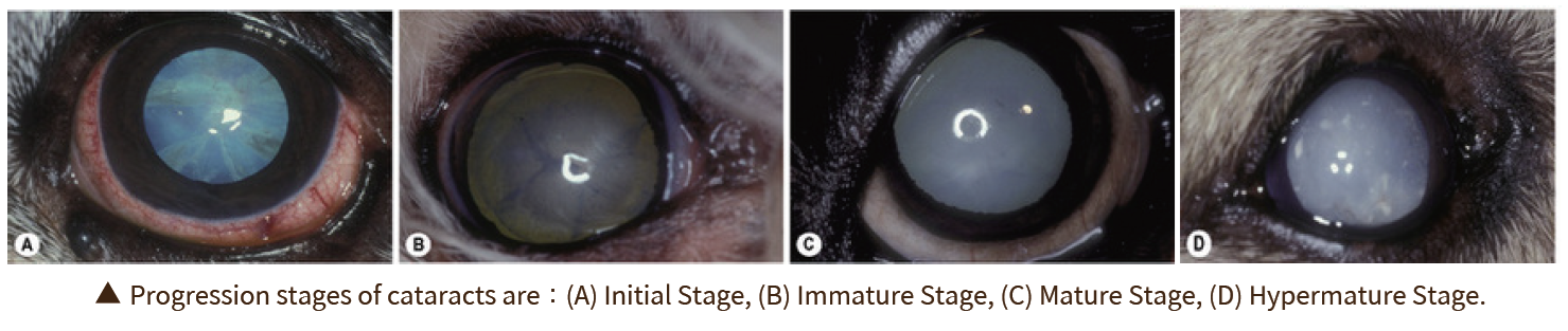
Cataracts can be classified into four stages based on the degree of lens opacity:
- Initial Stage: A distinct opaque white spot appears in the center of the pupil, but vision remains unaffected.
- Immature Stage: The lens begins to thicken both in the front and back, showing partial cloudiness. Vision becomes blurry, especially in low-light conditions, although some vision is still retained.
- Mature Stage: The entire lens becomes fully opaque and thickened, resulting in complete vision loss.
- Hypermature Stage: The clouded lens begins to shrink and clear up, making this stage prone to additional complications such as uveitis, glaucoma, and severe intraocular inflammation. This stage may also involve lens dislocation or fibrosis of the posterior capsule.
Impacts and Complications
The most noticeable early sign of cataracts is a change in eye color, where the lens begins to appear cloudy and white. It’s important to distinguish this from nuclear sclerosis, a common condition in older dogs. As the lens becomes cloudier, a dog’s vision deteriorates, leading to unintentional collisions with objects and increased sensitivity to the environment, often giving the dog a distant or blank stare.
Cataracts typically worsen over time, resulting in a range of eye-related complications. These can include increased sensitivity, lens-induced uveitis (LIU), glaucoma, and the rupture of zonular fibers around the lens, which may cause lens dislocation or subluxation, as well as opacification of the posterior capsule. As cataracts progress to the hypermature stage, additional complications such as intraocular bleeding, retinal detachment, and further instances of glaucoma may arise. Early diagnosis and treatment are essential for managing these complications effectively and improving the overall prognosis.
Diagnosis
A comprehensive ophthalmological examination is essential to assess the severity of cataracts and identify any associated complications. This examination typically includes tests for vision, pupillary light reflex, cataract severity and grading, and checks for any concurrent eye diseases or complications. Additional tests may include intraocular pressure measurement, tear production evaluation, corneal health assessment, fundus reflex and retinal examination, and ocular ultrasound.
Genetic testing can further confirm the presence of genetic defects and is valuable as a pre-breeding health check to prevent the transmission of hereditary cataracts to offspring.
References
Mellersh CS, McLaughlin B, Ahonen S, Pettitt L, Lohi H, Barnett KC. Mutation in HSF4 is associated with hereditary cataract in the Australian Shepherd. Vet Ophthalmol. 2009 Nov-Dec; 12(6):372-8.
Mellersh CS, Pettitt L, Forman OP, Vaudin M, Barnett KC. Identification of mutations in HSF4 in dogs of three different breeds with hereditary cataracts. Vet Ophthalmol. 2006 Sep-Oct; 9(5):369-78.
