Bioguard Corporation
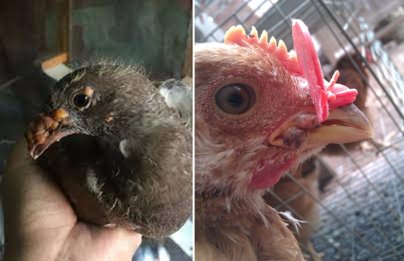
Fowl pox is a slow-spreading viral disease affecting chickens, turkeys, and various other birds. It is characterized by proliferative skin lesions that develop into thick scabs (cutaneous form) and lesions in the upper respiratory and digestive tracts (diphtheritic form). A presumptive diagnosis can be made in the field based on distinctive skin lesions, but confirmation requires detecting cytoplasmic inclusion bodies in affected cells.

Etiology
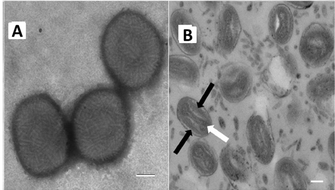
Fowl pox has a world-wide distribution and is caused by a DNA virus of the genus Avipoxvirus of the family Poxviridae. Virions are somewhat pleomorphic, generally brick-shaped (220–450 nm long×140–260 nm wide×140–260 nm thick) with a lipoprotein surface membrane displaying tubular or globular units (10–40 nm). The large DNA virus is resistant and may survive in the environment for extended periods in dried scabs.
Avipoxvirus affecting more than 230 species in 23 orders of wild and domesticated birds. Within the Avipoxvirus genus there are currently 10 recognized species: : Fowlpox virus, Canarypox virus, Juncopox virus, Mynahpox virus, Psittacinepox virus, Sparrowpox virus, Starlingpox virus, Pigeonpox virus, Turkeypox virus, and Quailpox virus, according to the International Committee on Taxonomy of Viruses.

Transmission
The fowl pox virus is abundant in lesions and typically spreads through contact with skin abrasions. Scabs shed from recovering birds in poultry houses can become airborne, leading to infection. Mosquitoes and other biting insects can also act as mechanical vectors, rapidly spreading the virus when mosquitoes are prevalent. The virus is highly resistant in dry scabs, making it easy to transmit to uninfected birds. Fowl pox can affect birds of any age and may occur year-round.
Clinical signs
Clinical signs are somewhat variable depending on the host species, virulence of the virus strain, distribution of lesions, and other complicating factors. In chickens and turkeys, signs may vary with two overlapping forms of the disease:
Cutaneous (dry pox): The dry form begins with a pimple or scab on non-feathered areas of the skin such as the comb, wattles, eyelids, feet, and legs. Eventually, the infection may spread to other feathered areas of the body. Infected birds often have difficulty eating and reduced feed intake and weight loss is common. Cutaneous lesions on the eyelids may cause complete closure of one or both eyes.
Diphtheritic (wet pox): The wet form produces diphtheritic, yellow canker lesions on oral mucous membranes, tongue, esophagus, or trachea. Lesions in the upper digestive and respiratory tract may result in inappetence and dyspnea, respectively. Other mild to severe respiratory signs may also occur. Lesions in the eye and nasal cavity lead to ocular or nasal discharges.
Diagnosis:
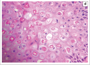
A presumptive diagnosis can be made in the field based on characteristic skin lesions. The diagnosis should be confirmed by laboratory testing. Microscopic examination of affected tissue sections stained with H&E reveals eosinophilic cytoplasmic inclusion bodies. Inclusions are also detected by fluorescent antibody and immunoperoxidase methods. Viral particles, with typical poxvirus morphology, can be detected by electron microscopy. The virus can be isolated in the chorioallantoic membrane (CAM) of chicken embryos, susceptible birds, or avian cell cultures. Field viruses can be detected in the laboratory by polymerase chain reaction (PCR).

Prevention and Control
Fowl pox and pigeon pox live virus vaccines are commonly used for immunization of chickens. Vaccination effectively prevents the disease and may limit spread within actively infected flocks. No treatment exists for birds infected with avian pox viruses.
References
Giotis ES, Skinner MA. Spotlight on avian pathology: fowlpox virus. Avian Pathol. 2019 Apr;48(2):87-90.
Molini U, Mutjavikua V, de Villiers M et al. Molecular characterization of avipoxviruses circulating in Windhoek district, Namibia 2021. J Vet Med Sci. 2022 May 25;84(5):707-711.

