Support

Search

Pancreatitis in Dogs

Dr. Sushant Sadotra Canine Pancreatitis is one of the most common endocrine diseases occurring in dogs. However, it is more prevalent in dog breeds such as Miniature Schnauzers, Yorkshire Terriers, Cocker Spaniels, Dachshunds, Poodles, and sled dogs. Pancreatitis, a severe inflammatory condition of the pancreas, can be short-term or long-term, based on the level of pancreatic tissue damage. It can be of acute or chronic type. It can be related to various clinical or subclinical signs and potentially life-threatening.  Causes: Pathogenesis In the initial stages of Pancreatitis, the pancreatic juice is secreted in lesser amounts. Inside the pancreas, a series of steps lead to the activation of pancreatic enzymes. Co-localization of zymogen granules and lysosomes activates trypsinogen to trypsin, which further activates other zymogens. Premature activation of these digestive enzymes causes local damage such as edema, bleeding, inflammation, and necrosis of the pancreas. The inflammation process invites WBCs to the site and increases cytokine production. Altogether, this will cause further damage to pancreases and other distant complications such as generalized inflammation, disseminated intravascular coagulation, disseminated lipodystrophy, hypotension, renal failure, pulmonary failure, myocarditis, etc. Clinical Findings: Some of the most common symptoms in dogs are: The milder form of Pancreatitis can be related to subclinical or may have minor or nonspecific signs of anorexia, lethargy, or diarrhea. Diagnosis: Among all the methods discussed, histopathology is the gold standard for the diagnosis of canine Pancreatitis. However, a combination of mentioned techniques can be implemented in clinical practice for the most reliable and accurate diagnosis. Treatment: Careful monitoring and supportive veterinary care should be given in acute cases to prevent systemic complications. If a dog with chronic pancreatitis has no sign of improvement, additional trial therapy with an immunosuppressive agent such as prednisone, prednisolone, or cyclosporine may be prescribed for the treatment. Treatment for chronic pancreatitis is challenging because of systemic complications such as hypothermia, acidosis, hypocalcemia, and single- or multiple-organ failure. Reference: Watson, P. (2015), Pancreatitis in dogs and cats: definitions and pathophysiology. J Small Anim Pract, 56: 3-12. Whitley EM. Comparative Pancreatic Pathology. Pathobiology of Human Disease. 2014:1101–23. doi: 10.1016/B978-0-12-386456-7.03415-8. Epub 2014 Aug 21. PMCID: PMC7149520. Xenoulis, P.G. (2015), diagnosis of pancreatitis in dogs and cats. J Small Anim Pract, 56: 13-26.

Kidney Diseases in Cats

Lloyd Alexandria Chavez, R.M.T   Cats possess a pair of kidneys located on either side of their abdomen, playing a crucial role in eliminating waste from their system. These organs are also key in regulating the balance of fluids, minerals, and electrolytes in the body, conserving water and protein, and supporting blood pressure and the production of red blood cells through the production of the hormone erythropoietin. Kidney disease in cats can manifest in several forms and can stem from various causes, typically classified into either acute or chronic categories. Acute kidney injury occurs when the kidneys are suddenly damaged, potentially impairing their function. This condition can affect both pets and humans and may result in diminished kidney performance. Fortunately, acute kidney injury can often be reversible, with approximately half of those affected—be they pets or humans—able to recover. The kidneys have a remarkable capacity for self-repair, provided the initial cause of injury is addressed and any exacerbating factors are eliminated. Recovery prospects depend on several factors, including the cause of the injury, its severity, whether other bodily systems are involved, the availability of treatments such as hemodialysis, and adherence to medical guidance.Chronic kidney disease (CKD), on the other hand, is frequently diagnosed in older cats and is characterized by a progressive decline in kidney function. Symptoms of CKD in cats include increased thirst and urination, reduced appetite, weight loss, vomiting, and a dull coat. CKD is an incurable condition that worsens over time, resulting from gradual kidney damage over months or years. Its progression and symptom onset are more gradual compared to acute kidney disease, which can emerge rapidly following significant kidney damage from causes such as infections or toxic substances like antifreeze or lilies. Cats with acute kidney disease typically exhibit severe symptoms swiftly. SYMPTOMS In the initial stages of chronic kidney disease (CKD), there may be no noticeable symptoms as the kidneys are still able to function adequately. However, as the disease progresses and the kidneys become increasingly impaired, symptoms will begin to manifest. These symptoms can develop gradually and may be subtle, making them difficult to detect. They include: DIAGNOSIS Chronic kidney disease is diagnosed through a series of blood and urine tests that measure levels of specific substances indicative of kidney function. These tests not only confirm the presence of kidney issues but also aid veterinarians in staging the disease, which helps in understanding its severity. The key laboratory tests include: Together, these tests provide a comprehensive assessment of kidney health, allowing for accurate diagnosis and staging of chronic kidney disease in cats. TREATMENT While it’s not possible to cure chronic kidney disease (CKD) or undo the damage that has occurred, steps can be taken to slow its progression and alleviate the symptoms. In cases where a cat is diagnosed with CKD and is showing signs of illness, the initial course of treatment may include: This treatment regimen aims to manage the symptoms and complications associated with CKD, improving the quality of life for cats living with this condition. Sources: https://www.pdsa.org.uk/pet-help-and-advice/pet-health-hub/conditions/chronic-kidney-disease-in-cats#:~:text=FAQs-,Overview,vomiting%2C%20and%20poor%20coat%20condition. https://www.petmd.com/cat/conditions/urinary/kidney-disease-cats https://www.ncbi.nlm.nih.gov/pmc/articles/PMC7379052/

C-Reactive Protein

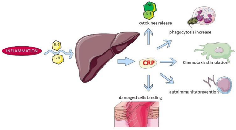
Sushant Sadotra The acute phase response (APR) is the early typical systemic response prompted by homeostasis disturbances such as injuries, infection, neoplasia, and other pathologies. Acute phase proteins get released from the liver into the blood after such stimulations. Therefore, elevated levels of such proteins can be used to indicate systemic inflammation. C-reactive protein, also called CRP, is an acute-phase protein synthesized in the liver and acts as a marker of the innate immune system response during inflammatory reactions. It is a 120 kDa composed of five identical subunits to form a pentamer structure. In Dogs, proinflammatory cytokines cause a rapid rise in the blood CRP concentration that also returns to normal without the provocative cause. Therefore it serves as an indicator and a crucial biomarker of inflammation. Besides projecting the degree of inflammation, CRP is helpful as a marker to examine the severity of a specific disease, such as gastrointestinal issues or diseases of other organs. Some of the biological functions of CRP are mentioned below: Factors inducing acute phase response, including CRP: CRP has been widely used in Europe and Japan to examine dogs with gastrointestinal and nongastrointestinal disorders. Some of the pathophysiologic conditions where CRP is frequently used as a biomarker are: CRP is a marker for chronic gastrointestinal signs, mucosal inflammation, systemic or gastrointestinal diseases, etc. Measurement of CRP and pancreatic lipase provide more sensitive and specific results. CRP can provide predictive information in dogs with CPV-2 infection. Dogs with hepatic disease have shown evaluated serum CRP levels. CRP is also the indicator of an acute abdomen in dogs with symptoms of abdominal pain, gastric dilatation or volvulus, small bowel obstruction, and peritonitis. CRP concentrations are higher in dogs with PLE, a disorder where excessive protein loss occurs through gastrointestinal mucosa. Dogs with Immune-mediated hemolytic anemia have high CRP levels. Tumor-associated inflammation or cytokine production leads to higher CRP concentrations in dogs with cancer of hematopoietic origins, such as lymphoma and leukemia. Measurement of canine CRP: POCTs track the inflammatory response in dogs as a rapid ancillary test or emergency. Evaluating CRP levels can be tested using available POCTs like the Point Strip canine CRP assay, LifeAssays canine CRP test, EURO-Lyser solo CRP test, and TECOdogCRP-quant. This commercially available test can estimate the scattering of light from a laser interacting with the CRP and anti-canine CRP antibody complex. This test is developed as Laser CRP-2 by Arrows Co., Ltd., Osaka, Japan. Many laboratories can estimate CRP at a large scale using commercially available multiple canine-specific CRP ELISA Tests such as Phase Range canine CRP ELISA37 (developed by Tridelta Development Ltd, Kildare, UK) and test invented by Waritani et al. Measurement of CRP in dogs can also be done through many automated immunoturbidimetric assays like the Turbovet canine CRP assay by Acuvet Biotech, Zaragoza, Spain; the Gentian canine CRP assay by Gentian AS, Moss, Norway; and TR-IMFA developed by Parra et al. Reference:

Rabbit Hemorrhagic Disease

Long Pham Introduction     Rabbit hemorrhagic disease (RHD) is a highly contagious and lethal viral disease caused by a virus from the Caliciviridae family. This disease seems to only affect European rabbits (Oryctolagus cuniculus),both domesticated and wild rabbits. However, a newer strain of the virus, RHDV2, can affect rabbits with previous immunity to RHD and also hares (Lepus spp.). The first outbreak of rabbit hemorrhagic disease was noted in China from a shipment of Angora rabbits from Germany in 1984 (1). The devastating nature of this disease can be seen as about 140 million domestic rabbits were killed in China during this outbreak in just less than a year (1). The next country to have an RHD outbreak was Korea due to importing rabbit fur from China (2). In 1986, the disease arrived in Italy and then spread to the rest of Europe (3). In 1988, the first outbreak in the Americas was reported in Mexico from imported rabbit products coming from China (4). However, Mexico was able to control RHD and had its last outbreak in 1992 (4). While RHD has been seen as a devastating disease that causes ecological and economical impacts, for New Zealand and Australia, RHD was considered as a method of pest control for the wild rabbits (5). Rabbit hemorrhagic disease virus was released in Wardang Island in South Australia, but in 1995, RHDV escaped and reach Australia (6). At the moment, RHD generally occurs in areas where European rabbits are found in the wild, with deadly outbreaks that can occur on almost all continents. Clinical signs Rabbit hemorrhagic disease virus has an incubation period of around 1-3 days. The virus can affect many internal organs (mainly targets the spleen, liver, and lungs), causing internal bleeding in infected rabbits. This disease can be present in 3 different forms: peracute, acute, subacute. Peracute form: Infected rabbit dies suddenly without showing any signs. Can be seen functioning normally before suddenly dying. Acute form: Rabbits can have a lack of motivation to move, ataxia, convulsions, subconjunctival hemorrhage, respiratory issues, and nosebleeds (Figure 1). For these infected rabbits, death can occur within 72 hours. Subacute form: These rabbits can have the same symptoms, but in a milder form. In this form, liver damage can occur, causing jaundice, with death that can occur weeks later. In general, most rabbits survive and develop some immunity to the virus. In general, rabbits that have clinical symptoms will die from the disease, while rabbits with the subacute form may survive. Rabbits that recover from the subacute form may have extensive liver damage, resulting in chronic liver diseases. Diagnosis Rabbit hemorrhagic disease can be confirmed through a post-mortem examination for the presence of an enlarged and pale liver. Molecular testing of live samples with PCR or ELISA can also confirm diagnosis. However, some tests may not be able to distinguish between RHDV and RHDV2. Figure 1: Rabbits with RHD often die suddenly and have bleeding from the nose due to internal bleeding. (Photo Credit: Dr. J.P. Teifke from theFederal Research Institute for Animal Health in Riems, Germany) Treatment and Disease Control         Rabbit hemorrhagic disease have several possible routes of transmission: nasal, oral, conjunctival, and through other places because hematophagous insects have been seen to be quite effective in spreading diseases (7). Rabbit hemorrhagic disease virus may be shed through the rabbit’s excretions and secretions, making direct contact with or contact with contaminated environment the likely method of transmission. The remains of infected rabbits may also be a source of infections and spreading of the virus because it is very resilient and has been seen to still be viable even after three months (8). Flies and birds can also be effective carriers of the virus because they can travel great distances with the resilient virus and contaminate the rabbit’s food source and habitat. Carnivores and scavengers are other possible carriers because the virus from consumed infected rabbits can transported and spread through the feces.      Currently, there is no cure for rabbit hemorrhagic disease. Supportive care may help, but it’s best to have preventive care with vaccinations. Hyperimmune antiserum can be effective if the rabbit has not showed clinical signs, but will be ineffective after the signs appear. For domestic rabbits, vaccinations is an effective preventative measure. However, for wild rabbits, the sheer number of rabbits would make vaccinations economically unfeasible at the moment.         To help prevent outbreaks, countries can place bans or restrictions on importing rabbit products, such as meats or furs, from other countries where rabbit hemorrhagic disease is endemic. Other preventative methods are culling, monitoring, following strict quarantining rules, and thoroughly disinfecting contaminated areas before bringing rabbits there again. Disinfection of the rabbit hemorrhagic disease virus can be done with a 4-10% solution of sodium hydroxide or 1-2% formalin solution. A 1:10 dilution of household bleach can also be used for disinfecting infected surfaces. However, since the virus is nonenveloped, an alcohol-based disinfection is not effective. Reference 1. Liu, S. J., H. P. Xue, B. Q. Pu, and N. H. Qian. 1984. A new viral disease in rabbits. Animal Husbandry and Veterinary Medicine (Xumu yu Shouyi) 16:253–255. 2. Park, N.Y., C. Y. Chong, J. H. Kim, S. M. Cho, Y. H. Cha, B. T. Jung, D. S. Kim, J. B. Yoon, J. Y. Park, and S. H. Wee, 1987. An outbreak of viral haemorrhagic pneumonia (tentative name) of rabbits in Korea. Journal of the Korean Veterinary Medical Association: 23: 603-610. 3. Cancellotti, F. M., and M. Renzi. 1991. Epidemiology and current situation of viral haemorrhagic disease of rabbits and the European brown hare syndrome. Revue Scientifique et Technique de l’OIE 10:409–422. 4. Gregg, D. A., C. House, and M. Berninger. 1991. Viral haemorrhagic disease of rabbits in Mexico: Epidemiology and viral characterization. Revue Scientifique et Technique de l’OIE 10:435–451. 5. Cooke, B. D. 2002. Rabbit haemorrhagic disease: Field Epidemiology and the management of wild rabbit populations. Revue Scientifique et Technique de l’OIE 21:347–358. 6. Cooke, B. D., and F. Fenner. 2002. Rabbit

Inflammatory Bowel Disease in Cats

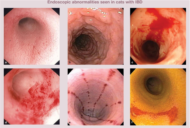
Sushant Sadotra Feline inflammatory bowel disease (IBD) is a chronic condition that affects a cat’s gastrointestinal (GI) tract. The walls of the GI tract become thickened due to the infiltration of inflammatory cells, disrupting the cat’s ability to digest and absorb food properly. Although IBD can affect cats of any age, middle-aged and older cats are more prone to it. The exact cause of IBD remains unknown, but current evidence suggests that it may result from an abnormal interaction between the immune system, diet, bacterial populations in the intestines, and other environmental factors. Genetic abnormalities in the immune system are also believed to play a role in feline IBD, based on similarities to IBD in humans and dogs. Depending on the region of the GI tract and the type of inflammatory cells involved, IBD can manifest in different forms. If the stomach is inflamed, it is called gastritis; if the small intestine is inflamed, it is called enteritis, and if the colon is inflamed, it is called colitis. Lymphocytic plasmacytic enteritis is the most common form of IBD, where inflammatory lymphocytes and plasma cells attack the small intestine. Eosinophils, another type of inflammatory white blood cell, may also be involved in feline IBD, but are usually part of a mixed population of inflammatory cells. Neutrophilic IBD, which involves neutrophils, and granulomatous IBD, which involves macrophages, are two less common forms of IBD. In some cases, IBD can cause inflammation of other abdominal organs, such as the liver and pancreas. It is important to identify the type of IBD affecting a cat through appropriate diagnostic procedures to provide the best possible treatment and management. Clinical symptoms: Feline IBD is often characterized by a set of common clinical signs that include weight loss, vomiting, decreased appetite, diarrhea, lethargy, and bloody stools. The severity and frequency of these signs can vary depending on which parts of the gastrointestinal tract are inflamed. For instance, if the inflammation is situated in the stomach or higher regions of the small intestine, the cat may experience chronic vomiting. Conversely, inflammation in the colon is more likely to cause diarrhea, with or without blood in the stool. Diagnosis When it comes to making a diagnosis of feline IBD, it is important to conduct a thorough workup since many of the symptoms of IBD can overlap with other diseases. To determine the root cause of the symptoms, your veterinarian will most likely recommend conducting baseline blood work, fecal examinations, X-rays, or an abdominal ultrasound to check for metabolic disease, feline leukemia, parasitic or bacterial infections, hyperthyroidism, and certain types of cancer. Intestinal lymphoma, a form of cancer, can be particularly challenging to distinguish from IBD in cats. Additionally, a veterinarian may also measure the levels of B vitamins B12 and folate in the bloodstream, as IBD can hinder the absorption of these vitamins from the GI tract. Finally, a hypoallergenic food trial may be conducted to rule out food allergy as a possible cause. To further diagnose feline IBD, a biopsy is required to evaluate the tissue under a microscope. Increased numbers of inflammatory cells in the intestinal wall indicate the presence of IBD. Endoscopy and surgery are two methods of performing gastrointestinal biopsies, both of which require general anesthesia. However, surgery may be recommended for patients with suspected liver or pancreatic disease to biopsy these organs as well. Treatment If you suspect that your furry friend has intestinal parasites, it is important to consult with a veterinarian who will recommend appropriate treatment. The initial steps usually involve a combination of dietary changes and medications. Since there is no one-size-fits-all solution, your vet may need to experiment with different diet and medication combinations to determine the best therapy for your pet. Dietary Management If your cat is suffering from Inflammatory Bowel Disease (IBD), it is likely that dietary allergens are playing a role. Your veterinarian may suggest undergoing a food trial using hypoallergenic diets to help alleviate the symptoms. These diets include protein or carbohydrate sources that your cat has never consumed before. Some common initial choices are diets based on rabbit, duck, or venison. In case the symptoms do not improve with a hypoallergenic diet, your cat may benefit from diets that are high in fiber, low in fat, and easily digestible. It is important to note that it may take several weeks or even longer for your cat to show signs of improvement after a diet change. During any food trial, it is crucial to eliminate all other food sources, including table scraps, flavored medications, and treats. Medical Treatment Reference

Canine Chronic Hepatitis

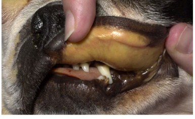
Chavezlloyd Alexandria Baldago Chronic hepatitis is a condition in dogs that may occur due to various disease processes. It indicates a previous occurrence of inflammation and possibly cell death in the liver. The inflammation is caused by the infiltration of different types of white blood cells that are involved in the immune system. Necrosis, which refers to the death of a large number of liver cells, may also occur. The invasion of white blood cells and cell death in a dog’s liver can be due to previous damage caused by infectious agents like viruses or bacteria or as a result of toxic damage. Toxic damage may occur due to poisons ingested by the dog or abnormal accumulation of substances required by the body, such as copper. Though some breeds like Bedlington Terriers, Labrador Retrievers, Doberman Pinschers, Dalmatians, West Highland White Terriers, Welsh Corgis, Keeshonds, and others are notably affected, this toxicity can affect any breed of dog. Copper accumulation in hepatocytes can cause oxidative damage, and its presence can escalate hepatocyte damage caused by other factors. Until dietary recommendations are modified and implemented, this problem will continue to affect canines. Inflammation and cell death can also occur due to a primary attack by the immune system against liver cells, known as an “autoimmune” disease. Cancer in the liver is not called chronic hepatitis, even if it causes similar damage.   Chronic vs Acute The term “chronic” refers to a condition that has been causing damage for a prolonged period, typically lasting several weeks or more. On the other hand, “acute” hepatitis is usually characterized by a shorter duration of just a few days. While some cases of acute hepatitis can be treated successfully, many types of chronic hepatitis are not curable. However, with appropriate treatment and close monitoring, a significant number of patients with chronic hepatitis can maintain a good quality of life with minimal clinical symptoms for an extended period. Chronic hepatitis can affect any breed of dog regardless of gender or age, although it is more common in middle-aged or older dogs. Certain breeds may be more prone to specific types of hepatitis. For instance, some breeds may develop chronic hepatitis due to the accumulation of copper in the liver cells. The excessive concentration of copper harms the liver cells and, if left untreated, typically leads to severe chronic hepatitis. Symptoms The liver has multiple functions, therefore the clinical signs associated with liver disease can vary significantly. Symptoms of hepatitis in dogs can include: Lack of appetite Lethargy Vomiting Increased urination Excessive thirst and urination Swollen belly filled with fluid (ascites) Yellowish gums (jaundiced) and moist tissues Seizures, mental dullness Diagnosis Apart from obtaining a detailed history of the affected dog’s health prior to the appearance of symptoms, the next step would be to conduct a comprehensive physical examination of the dog, which includes carrying out a blood chemical profile, a complete blood count, an electrolyte panel, and a urinalysis. The results of the bloodwork will enable your veterinarian to identify any signs of impaired kidney function. To further evaluate the disease, complete abdominal ultrasonography is essential. This diagnostic tool screens for concurrent diseases and helps acquire bile. It’s important to note that the liver can appear normal upon examination, and changes seen in chronic hepatitis can include uniform increases in liver echogenicity, decreased distinction of portal vein margins, and normal to small liver size. Abnormalities in the liver parenchyma, biliary tree, portal vein, and peritoneum should be assessed, along with acquired shunting and the presence of free peritoneal fluid. In order to accurately diagnose chronic hepatitis, a liver biopsy is necessary. There are 3 ways to perform a liver biopsy, surgically, via laparoscopy, or through the skin using a special needle under ultrasound guidance. However, surgical or laparoscopic biopsies are more informative compared to ultrasound-guided biopsies. The information obtained from the biopsy is required to determine the type and severity of liver disease, as well as to assess your dog’s prognosis.   Treatment The treatment for chronic hepatitis in dogs is a complex process that depends on the severity and type of liver disease, as well as the clinical signs exhibited by the dog. In severe cases, hospitalization, intravenous fluid therapy, and supportive care may be required. The most common medications prescribed for treating the disease are immunosuppressive or anti-inflammatory. In some cases, dietary changes may be necessary. Certain medications may also be used in specific situations, such as dogs whose illness is linked to copper accumulation, abdominal fluid build-up, or neurological symptoms. Life Expectancy Although this condition cannot be cured with the available treatments, the good news is that the dog can still live a good quality of life for months and even years with continued therapy. Regular blood work is necessary to ensure the dog is responding well to the treatment. This helps to adjust the medication and keep the dog relatively free of clinical signs. References: https://bluepearlvet.com/medical-articles-for-pet-owners/chronic-hepatitis-in-dogs/ https://todaysveterinarypractice.com/hepatology/canine-chronic-hepatitis-diagnosis-treatment/ https://www.msdvetmanual.com/digestive-system/hepatic-diseases-of-small-animals/canine-chronic-hepatitis#:~:text=Canine%20chronic%20hepatitis%20is%20a,be%20predisposed%20to%20chronic%20hepatitis.

Respiratory Tract Disease Complex in Cats
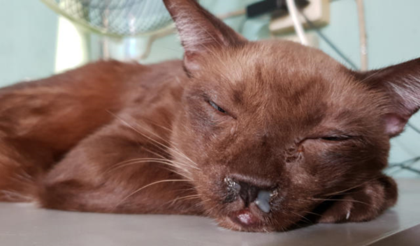
Sushant Sadotra, PhD/Diagnostic specialist Feline respiratory disease (FRD) syndrome or feline upper respiratory tract disease complex is a common infection in cats caused mainly by Feline Herpesvirus (FHV-1), Feline Calicivirus (FCV), Chlamydophila felis, Mycoplasma spp., and Bordetella bronchiseptica. About 90% of all upper respiratory infections are caused by FHV-1 and FCV. Common Symptoms: ·       Sneezing ·       Nasal congestion ·       Conjunctivitis (inflammation of the membranes lining the eyelids) ·       Discharge from the nose or eyes (clear, purulent, or cloudy containing pus). ·       Difficulty breathing ·       Ulcers in the mouth Less specific symptom ·       Less appetite ·       Lethargy ·       Fever ·       Enlarged lymph nodes ·      Blepharospasm (squinting) Sources of infection: ·       Susceptible cats can get an infection by contagious particles in saliva or secretions from the nose or eyes shredded by an infected cat. ·       Most cases are associated with direct contact ·       Natural transmissions can also occur via aerosol droplets. Stress may also cause a secondary course of illness. Real-Time PCR for Diagnosis: A definitive diagnosis is based on clinical signs and laboratory testing for the isolation and identification of the infection. The Polymerase chain reaction (PCR) test is one of the sensitive tests and most reliable for detecting the presence of infectious agents. PCR detects the genomic material of the pathogen and determines its presence in the host. It is often more sensitive and specific than other available tests. However, false negative results are still expected. In the case of patients with latent herpes infections where the FHV-1 is found in the trigeminal ganglion can give negative PCR results. In the case of Chlamydophila, 2-3 days of antibiotic treatment for patients can also have negative PCR results. Samples of ocular, nasal, or caudal pharyngeal secretions for PCR assay are best for the diagnosis and identification of causative agents. Sample collection tips: ·       Ocular: Moisten with tears/exudate well or firmly swab both of the conjunctival sacs with a sterile swab. ·       Clinical lesions: Prefer to swab from the nasal and pharyngeal areas. After sample collection, place the swab into the preservation buffer and mix thoroughly. If not for immediately use, please keep the mixture at 4°C (no more than 3 days) until nucleic acid extraction.