Spinal Muscular Atrophy in Cats
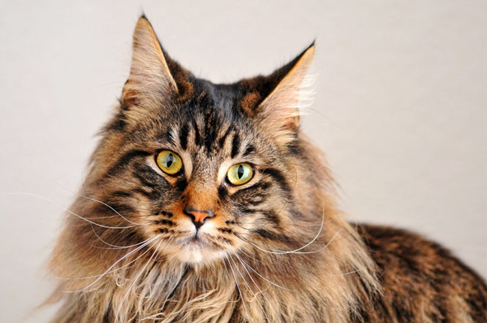
Bioguard Corporation Spinal muscular atrophy (SMA) an autosomally recessive inherited neurodegenerative disorder seen in Maine Coon cats. The disease is characterized by weakness and atrophy in muscles due to loss of motor neurons that control muscle movement. Affected cats first show signs of disease around 3–4 months of age. Clinical signs include tremors, abnormal posture, and weakness in muscle. Pathogenesis SMA in Maine Coon cat is caused by the deletion of a 140 kb LIX1 gene on the A1q chromosome. Although the function of LIX1 is not yet clear, it is presumed to be related to RNA metabolism. The LIX1 is highly expressed in the central nervous system, primarily in spinal motor neurons, thus offering explanation of the restriction of their function in case of feline SMA. The disease is inherited as an autosomal recessive. Clinical signs In the earliest stages of SMA, a vet or other professional will be able to notice a slight weakness in the affected kittens. Subtle tremors in a kitten’s hind legs can also be an early symptom of SMA. Within 1 to 2 months after the symptoms appear, the muscles will gradually atrophy. After that, the symptoms will gradually become serious, the movements will become clumsy, the agility will be lost in jumping, and even breathing symptoms will appear. Muscle atrophy affects only the hind legs, and some severely affected cats move by crawling on the front legs, although mental status is not affected. The condition slowly stabilizes after up to 8 months, and some cats with SMA have more or less severe symptoms for up to 9 years. Diagnosis The first step is a general physical exam. Your doctor will look for muscle weakness and any other signs of SMA. Serum creatine kinase (creatine) elevations and electrical findings in cats are very similar to mild spinal muscular atrophy in humans. Examination of the muscles reveals neurogenic atrophy, and examination of the central and peripheral nervous systems will reveal loss of anterior horn cells. To ascertain if a cat has SMA, the diagnostic approach encompasses a physical examination, electromyography, muscle biopsy, testing for serum creatine kinase levels, and genetic testing, all aimed at facilitating early identification and intervention. Since SMA is common in Maine Coons, it is recommended that Maine Coons with similar symptoms or those intended for breeding undergo this test. References Fyfe JC, Menotti-Raymond M, David VA, et al. An approximately 140-kb deletion associated with feline spinal muscular atrophy implies an essential LIX1 function for motor neuron survival. Genome Res. 2006 Sep;16(9):1084-90.
Polycystic Kidney Disease in Cats
Bioguard Corporation Polycystic kidney disease (PKD) is a chromosomally dominant genetic disorder; it can occur in humans, cats, dogs, and other animals. In the renal cortex and medulla, there are cysts of various sizes and fluid-filled, so it is commonly known as the bubble kidney. Cysts increase in size and number over time, replacing kidney tissue and affecting their ability to filter waste from the blood, leading to chronic kidney failure. Pathogenesis Polycystic kidney disease is primarily caused by point mutations in the PKD1 gene, which is inherited as a dominant genetic disorder. The PKD1 gene plays a crucial role in regulating polycystin, a protein found on the cell membrane. A deficiency in PKD1 leads to underdevelopment of the renal tubules and collecting ducts in the renal cortex and medulla, preventing proper drainage of urine filtered by the renal glomeruli, which results in the formation of cysts characteristic of polycystic kidney disease. In the liver, this condition can also cause significant enlargement of bile ducts near the portal vein and may lead to bile duct fibrosis. Clinical symptoms Cats with polycystic kidney disease (PKD) are born with abnormal kidneys, though symptoms typically do not manifest until they are between 3 and 10 years old, with an average onset around 7 years. While the disease is present from birth, there are no noticeable symptoms in its early stages. Symptoms appear only when the disease progresses to a point where kidney tissue necrosis and kidney failure occur. As renal cysts enlarge over time, they compress the renal parenchyma, leading to irreversible kidney failure. Affected cats may experience a decreased appetite, weight loss, depression, and lack of energy. Clinical symptoms include increased thirst (polydipsia), frequent urination (polyuria), anorexia, vomiting, lethargy, and muscle twitching in the abdominal area. In severe cases, movement disorders (ataxia) or neurological issues may arise. Blood tests may reveal elevated blood urea nitrogen (BUN) and creatinine (CRE) levels, anemia, and high blood pressure. Early detection At present, it is possible to know whether cats have polycystic kidney disease through ultrasound testing and genetic testing. At 16 weeks of age, about 75% of cats with this problem had cyst-like structures on ultrasound scans, and by 36 weeks of age, 91% of cats had cysts. The accuracy of such a structure increases with age; generally speaking, when cats are over 10 weeks old, when using ultrasonic scanning, the accuracy can reach 90~95%. Genetic testing, which refers to the detection of its genotype, will be 100% accurate and can be performed at any age Breed predisposition Polycystic kidney disease mainly occurs in long-haired cats, and studies have shown that up to 38% of Persian cats have an abnormal PKD1 gene. Mainly affects cats of Persian and Persian-related breeds, such as Chinchillas, but other breeds such as Ragdolls, Scottish Folds, or other shorthair breeds such as Himalayans and Exotics have also been reported It is possible to have this genetic disorder. In addition, Meeks’s condition is relatively rare. References Schirrer L, Marín-García PJ, Llobat L. Feline Polycystic Kidney Disease: An Update. Vet Sci. 2021 Nov 8;8(11):269.
Feline Hypertrophic Cardiomyopathy
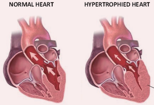
Bioguard Corporation Hypertrophic cardiomyopathy (HCM) is a primary, familial, and hereditary heart condition, and it is the most common heart disease in cats. Its key characteristic is primary concentric left ventricular hypertrophy (thickening of the heart wall), which occurs without pressure overload (such as from aortic stenosis), hormone stimulation (like in hyperthyroidism or acromegaly), myocardial involvement (such as from lymphoma), or other non-cardiac diseases. The heart consists of four chambers: the left atrium and ventricle, and the right atrium and ventricle. The right side of the heart pumps deoxygenated blood to the lungs, while the left side pumps oxygenated blood to the rest of the body. In hypertrophic cardiomyopathy (HCM), some cardiomyocytes are unable to function properly, causing the normal ones to enlarge in an attempt to maintain the heart’s output. However, this excessive thickening of the myocardium leads to a thickened left ventricle that encroaches on the ventricular space. As a result, the ventricle’s capacity to hold a normal amount of blood is reduced, and the myocardium becomes stiffer with decreased contractility. This alters the pressure within the left side of the heart, eventually causing the left atrium to enlarge. An enlarged left atrium increases the risk of congestive heart failure (CHF) in cats, which is marked by fluid accumulation in the lungs (pulmonary edema) or around the lungs (pleural effusion). HCM can occur at any age, but it is most common in adult cats around six years or older. Breeds such as Maine Coon, Ragdoll, and Domestic Shorthair are most frequently affected, while Persian, British Shorthair, and American Shorthair cats are also at higher risk. The exact cause of HCM in cats is not fully understood. Research suggests that mutations in the myosin binding protein C gene (MYBPC3) are linked to HCM in Maine Coon and Ragdoll cats. Specifically, the mutations A31P and R820W in the MYBPC3 gene are associated with this condition in these two breeds. The MYBPC3 mutation exhibits incomplete penetrance, meaning it is not a purely dominant trait. Cats with one copy of the mutated gene have a relative risk of HCM that is about 1.8 times higher than normal cats. However, cats with two copies of the mutation have a significantly higher relative risk, about 18 times greater. Despite this, some Maine Coon cats without the MYBPC3 mutation have also been diagnosed with HCM. In studies, the incidence of HCM in cats without the original mutant gene was approximately 5.4%. This indicates that while the MYBPC3 mutation is a significant factor, it is not the sole cause of HCM in Maine Coon cats, and other contributing factors remain unclear. Clinical Symptoms Most cats with HCM show no clinical symptoms, especially those with mild to moderate disease, making early detection challenging. Even severely affected cats may initially be asymptomatic but typically progress to left heart failure, systemic thromboembolism, or sudden death. Cats with heart failure exhibit signs such as shortness of breath and dyspnea due to pulmonary edema or pleural effusion. Systemic thromboembolism commonly presents as hind limb paresis or paralysis, accompanied by acute pain, lack of pulse, and fever. Genetic Testing This test is recommended for purebred cats with a genetic predisposition, especially Maine Coon and Ragdoll breeds.
GM1 Gangliosidosis in Cats
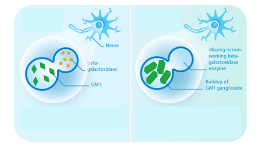
Bioguard Corporation GM1 gangliosidosis is a lysosomal storage disorder caused by deficiency of the enzyme β-galactosidase. Mutations in the GLB1 gene, encoding β-galactosidase, cause the progressive, neurosomatic, lysosomal storage disorder. Cats affected with gangliosidosis have progressive neurologic dysfunction around 3 months of age and premature death around one year old. Pathogenesis Gangliosides, normally hydrolyzed by β-galactosidase, are the main glycolipids of neuronal plasma membranes. Absent or reduced β-galactosidase activity leads to the accumulation of β-linked galactose-containing glycoconjugates including the glycosphingolipid (GSL) GM1-ganglioside in neuronal tissue. Instead of being broken down and recycled, this excess material is stored in membrane bound sacs (vacuoles) in cells. As the number of vacuoles increase, there are less available space and resources for normal cell function so that neuronal cell death and degeneration occurs, damaging the central nervous system and other organs. Clinical signs Kittens with GM1 gangliosidosis appear normal at birth and successfully achieve all developmental milestones. However, the accumulation of gangliosides in neuronal cells causes central nervous system damage. Clinical signs are apparent in cats at 2-3 months of age. Gangliosidosis is clinically characterized by discrete head and limb tremors and lack of coordination of movement. Other clinical signs include ataxia; tremor; visual disorder, which may have been due to cortical blindness; and seizures. The progressive accumulation of GM1 ganglioside in the central nervous system and cerebrospinal fluid ultimately proves fatal for cats at a young age. Diagnosis To rule other diseases causing symptoms similar to GM1 in cats, the diagnosis of gangliosidosis is carried out based on comprehensive findings using various types of specimens for histological, ultrastructural, biochemical and genetic analyses. A diagnosis of GM1 gangliosidosis requires biochemical identification of the storage product and enzyme deficiency. Affected animals had lower β-galactosidase activity in the liver and increased levels of GM1 gangliosides in the brain. In addition, DNA testing can be used to detect the mutation causing GM1 gangliosidosis in Korat cats; it can identify affected cats, carriers and normal cats, without the genetic mutation. Breeds such as the Siamese, Korat, Oriental Shorthair, Balinese, Havana Brown, Birman, Burmese, and Singapore Cat are more suscep-tible. It is recommended to conduct this test before breeding.
Cryptosporidiosis
Bioguard Corporation Cryptosporidiosis is an illness you get from the parasite Cryptosporidium. It causes watery diarrhea and other gastrointestinal (gut) symptoms. In addition to stomach infection, this parasite can infect the respiratory system causing a cough and/or problems breathing. The family Cryptosporididae belongs to the phylum Apicomplexa characterized by an anterior (or apical) polar complex (with apical rings, micronemes, and subpellicular microtubules), which allows penetration into host cells. Cryptosporidium species are able to infect a broad range of hosts including humans, domestic and wild animals (mammals, birds, fish, marsupials, reptiles, and amphibians) worldwide. Transmission and Life Cycle Humans and animals become infected with Cryptosporidium by touching anything that has come in contact with contaminated feces, although the most common mode of transmission is represented by ingestion of oocysts in contaminated food and water or air. Cryptosporidium has three developmental stages: meronts, gamonts, and oocysts. They reproduce within the intestinal epithelial cells. Two types of oocysts, thick-walled and thin-walled, are produced during sexual reproduction. Thick-walled oocysts are excreted from the host into the environment, whereas thin-walled oocysts are involved in the internal autoinfective cycle and are not recovered from stools. Oocysts are infectious upon excretion, thus enabling direct and immediate fecal-oral transmission. Clinical Symptoms The most common symptoms of cryptosporidiosis are watery diarrhea and stomach cramps. Other symptoms may include fever, nausea, vomiting, and loss of appetite. Symptoms and severity of infection vary with the age and immune status of the host. Cryptosporidium infections are uncommonly detected in cats and dogs. Cryptosporidiosis can sometimes make dogs and cats sick, but animals with signs are atypical. In most cases, epithelial damage is minimal, but in severe cases, infection is associated with losing the ability to maintain water balance. Clinical signs are usually restricted to mild diarrhea unless the host is immunosuppressed or has another underlying condition such as viral infection or malignancy. Diagnosis Cryptosporidiosis is a diarrheal disease that is spread through contact with the stool of an infected person or animal. The disease is diagnosed by examining stool samples. Oocyst excretion is intermittent, and multiple stool samples may be needed. Diagnostic methods include: Microscopic examination: Typically, stool samples are analyzed microscopically using various techniques, including acid-fast staining and Ziehl-Nielsen staining. Real-time PCR: The most accurate method for detecting Cryptosporidium spp. is through a fecal PCR assay. Immunologic tests: These include direct fluorescent antibody tests and enzyme immunoassays to detect Cryptosporidium sp. antigens. Treatment and Prevention Most patients with healthy immune systems will recover from cryptosporidiosis without treatment. Supportive measures, oral or parenteral rehydration, and hyperalimentation may be needed for immunocompromised patients with severe disease. The best way to prevent the spread of Cryptosporidium at home is by practicing good hygiene. References Sardinha-Silva A, Alves-Ferreira EVC, Grigg ME. Intestinal immune responses to commensal and pathogenic protozoa. Front Immunol. 2022 Sep 16;13:963723. Sponseller JK, Griffiths JK, Tzipori S. The evolution of respiratory Cryptosporidiosis: evidence for transmission by inhalation. Clin Microbiol Rev. 2014 Jul;27(3):575-86. Watier-Grillot S, Costa D, Petit C, et al. Cryptosporidiosis outbreaks linked to the public water supply in a military camp, France. PLoS Negl Trop Dis. 2022 Sep 12;16(9):e0010776.
Diagnosis of Feline Respiratory Mycoplasma Infection
Bioguard Corporation In cats, ’mucosal’ mycoplasma infections typically cause ocular and respiratory disease, and less frequently neurological or joint disease. These Mycoplasma species are distinct to the haemotropic mycoplasmas that target red blood cells, causing hemolytic anemia in cats. Mycoplasma felis is typically associated with Upper Respiratory Tract Disease (URTD) in cats. Transmission M. felis is mainly transmitted from an infected cat to an in-contact one by aerosol, but also by grooming. Stresses, including overcrowding environments, concurrent respiratory viral infections, and poor hygienic situations, may promote transmission of the infection between cats. Clinical symptoms Mycoplasma felis is typically associated with URTD but sometimes it may be associated with lower respiratory tract infections. Common clinical signs include clear or colored discharge from the eyes or nose, coughing, sneezing, conjunctivitis, chemosis, lethargy, and anorexia. Lower respiratory tract infections can result in pneumonia with fever, cough, tachypnoea, and lethargy. Diagnosis Culture of mycoplasmas can be used to demonstrate infection, but it takes time for culture and rapid transport of samples to the laboratory is required. Demonstration of organisms via real-time PCR is increasingly being used to circumvent the difficulties with culture, Treatment Antimicrobial therapy is commonly used to treat mycoplasma respiratory infections. Doxycycline is a good first line agent because it is well tolerated by cats and relatively narrow in spectrum. The recommended dose is 5 mg/kg, PO, q12h or 10 mg/kg, PO, q24 (Lappin et al., 2017). Oxytetracycline or chlortetracycline ophthalmic ointment can be used q6h in addition as topical treatment. References: Vekšins A. Feline upper respiratory tract disease – Computed tomography and laboratory diagnostic. Vet World. 2022 Jul;15(7):1880-1886. Framst I, Ramesh P, Cai HY, Maboni G. Complete genome sequences of Mycoplasma cynos and Mycoplasma felis isolated from dogs and cats with infectious respiratory disease. Microbiol Resour Announc. 2024 Apr 11;13(4):e0124323.
Tritrichomonas Infection in Cat
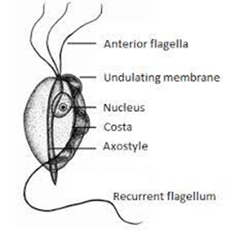
Bioguard Corporation Tritrichomonas foetus is a significant cause of large bowel diarrhea and persistent colitis in cats. These pear-shaped organisms have three anterior flagella and one posterior flagellum. They have a distinctive undulating membrane, which gives them a similar appearance to Giardia. However, they do not form cysts and are transmitted directly from one host to another as trophozoites. The infection is most prevalent among young cats living in close quarters, such as in densely populated catteries and shelters. A notable investigation into purebred show cats discovered a 31% infection rate among 117 cats spanning 89 catteries, as detailed in Gookin’s 2004 study Clinical Signs Some cats infected with T. foetus may not exhibit any symptoms, particularly older cats that are in good health. However, most cats with T. foetus infection suffer from mild to severe lymphoplasmacytic and neutrophilic colitis, which causes recurrent episodes of large bowel diarrhea that may vary in consistency from semiformed to “cow pie” and emit a foul odor. Diarrhea may contain fresh blood or mucus. Insevere cases, kittens may experience painful anal irritation, fecalincontinence, or even rectal prolapse. Affected cats usually maintain a healthy appearance and good body condition overall. The presence of diarrhea can be exacerbated by other intestinal infections or parasites, particularly Giardia and ryptosporidium. Diagnosis To confirm the infection of T. foetus, there are three methods available – direct fecal microscopy, fecal culture, and fecal polymerase chain reaction (PCR) assay. Direct fecal microscopy involves identifying the motile trophozoites T. foetus in fresh wet smears of diarrheic feces taken directly from the rectum. This method identifies the organisms in about 14% of cases and is less effective with formed or dried feces. In cats who have been treated with antibiotics recently, the detection rate decreases. Trichomonads, which resemble Giardia in size and shape, can be distinguished by their unique undulating membrane and rapid, jerky motility, contrasting with Giardia’s “falling leaf” movement. Fecal culture can be done in-house or at a specialized lab. It helps increase the chances of identifying the organisms. In specific cases, a saline flush might be performed by inserting a catheter through the cat’s anus to wash the colon with saline, followed by aspiration of fecal material. Fecal PCR is the most accurate method for identifying T. foetus. To perform this test, the fecal sample should not contain any litter. This technique detects the organism’s DNA traces in the cat’s stool. It’s best to conduct testing on cats that have not received antibiotics for at least two weeks for the most accurate test results. Antibiotics can temporarily reduce the number of T. foetus, leading to false negatives. Treatment Often, many approaches for treating chronic diarrhea have been tried unsuccessfully before a true diagnosis of T. foetus is confirmed. Tritrichomonas foetus is resistant to most antibiotics and is extremely difficult to eradicate (Gookin 2001). Numerous antibiotics have been evaluated. Some antibiotics reduce the number of organisms and improve the diarrhea without eliminating the infection, so diarrhea relapses whenever antibiotics are stopped. Diarrhea is typically refractory to corticosteroids. The most successful treatment for eliminating T. foetus is ronidazole (30 mg/kg PO, once or twice daily for 14 days). The side effects in some cats include lethargy, decreased appetite, and neurotoxicity. Cats with neurotoxic signs usually improve when the drug is stopped, but recovery can take 1 to 4 weeks. Ronidazole should not be used in pregnant and nursing queens or in very young kittens. Ronidazole is not approved for veterinary or human use, but some pharmacies compound chemical grade ronidazole for veterinary use. Because of its bitter taste, ronidazole compounded in gel caps is better tolerated than flavored suspension. When prescribing ronidazole obtain informed consent and instruct owners to wear protective gloves when handling it. Management of Tritrichomonas foetus Infection In cases where cats show mild or sporadic symptoms of diarrhea caused by T. foetus, and treatment is not possible due to potential side effects, costs, or the owner’s preferences, it is important to know that diarrhea may naturally go away with time, which can take up to two years. However, such cats are likely to remain lifelong carriers of the parasite. The outlook for cats receiving treatment is generally positive. A majority of treated cats exhibit better stool consistency in just a few days, though diarrhea might linger briefly as related secondary inflammation subsides. Nonetheless, around 25% of cases might experience a continuous infection despite initial treatment. Fortunately, T. foetus has a short lifespan outside its host and is easily neutralized by common disinfectants. To mitigate infection risks, it’s advised to uphold strict litter box cleanliness through daily cleansing, isolate cats under treatment, minimize stress factors, prevent overcrowding, and implement regular screenings in breeding and shelter settings whenever feasible.